Overview
This article presents a methodology for high-resolution fluorescent voltage-sensitive dye imaging to study neuronal activity in the crab stomatogastric ganglion. The technique allows for the optical recording of membrane potential changes and synaptic interactions among neurons.
Key Study Components
Area of Science
- Neuroscience
- Electrophysiology
- Imaging Techniques
Background
- Voltage-sensitive dyes can optically record changes in membrane potential.
- Central pattern generators in crustaceans are critical for understanding neural networks.
- Traditional recording techniques can be limited by electrode placement.
- This method aims to overcome those limitations.
Purpose of Study
- To develop a technique for recording neuronal activity optically.
- To analyze synaptic interactions in the stomatogastric ganglion.
- To provide insights into central pattern generator function.
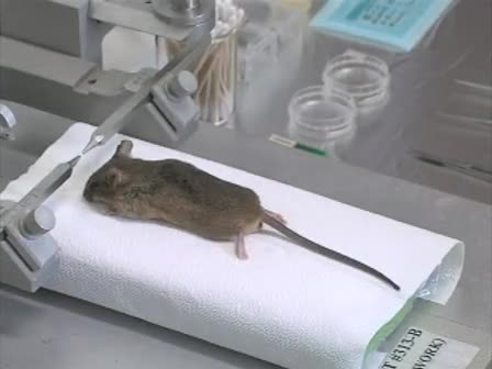
Methods Used
- Loading identified neurons with voltage-sensitive dyes via ion injection.
- Simultaneous extracellular recording of motor nerve activity.
- Analyzing fluorescence changes corresponding to membrane potential variations.
- Using imaging systems to visualize dye spread and neuronal activity.
Main Results
- The optical recording technique successfully captured synaptic interactions.
- Dye filling allowed for long-term recordings without damaging neurons.
- Results indicated the potential for this method to enhance understanding of neural networks.
Conclusions
- This technique provides a novel approach to studying neuronal dynamics.
- It can be applied to various biological systems beyond crustaceans.
- Further optimization is needed for those new to the method.
What are voltage-sensitive dyes?
Voltage-sensitive dyes are fluorescent compounds that change their fluorescence properties in response to changes in membrane potential.
How does this method compare to traditional recording techniques?
This method allows for optical recordings without the steric restrictions and potential damage associated with electrode placement.
What is the significance of studying the stomatogastric ganglion?
The stomatogastric ganglion serves as a model for understanding central pattern generators and neural network dynamics.
Can this technique be applied to other organisms?
Yes, the methodology can be adapted for use in other systems, including vertebrates.
What challenges might new users face with this technique?
New users may struggle with dye selection, injection preparation, and setting parameters for image data analysis.