Overview
This protocol describes the preparation and imaging of resin-embedded brain tissue in three dimensions using a focused ion beam scanning electron microscope. The method allows for high-resolution imaging of specific brain regions.
Key Study Components
Area of Science
- Neuroscience
- Imaging Techniques
- Histology
Background
- Focused ion beam scanning electron microscopy offers advantages over traditional imaging methods.
- It provides higher Z resolution and can image larger volumes of resin-embedded samples.
- This technique is particularly useful for studying brain tissue architecture.
- Serial imaging allows for comprehensive analysis of the region of interest.
Purpose of Study
- To collect serial images of brain tissue for detailed structural analysis.
- To demonstrate a method that improves imaging resolution and volume capability.
- To facilitate the study of specific brain regions in a three-dimensional context.
Methods Used
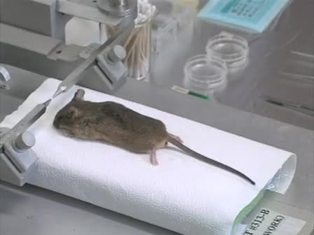
- Dissection and embedding of brain tissue in resin.
- Serial sectioning and staining of brain slices.
- Trimming and mounting of the resin block for imaging.
- Imaging using focused ion beam scanning electron microscopy.
Main Results
- Successful collection of serial images through the region of interest.
- High-resolution imaging achieved with isotropic voxels.
- Demonstrated capability to visualize structures from multiple angles.
- Results show significant improvement over traditional imaging methods.
Conclusions
- The protocol provides a reliable method for imaging resin-embedded brain tissue.
- Focused ion beam scanning electron microscopy is effective for detailed structural studies.
- This technique can enhance our understanding of brain tissue organization.
What is the main advantage of using focused ion beam scanning electron microscopy?
It achieves much higher Z resolution and can image larger volumes compared to traditional methods.
How are the brain samples prepared for imaging?
The samples are sectioned, stained, and embedded in resin before imaging.
What type of brain tissue is used in this protocol?
The protocol uses fixed brain tissue from anesthetized perfused rats.
Can this method be applied to other types of tissues?
While this protocol is specific to brain tissue, similar methods may be adapted for other types of resin-embedded samples.
What is the purpose of using a vibrato slice?
It allows for precise cutting of coronal sections of the brain tissue.
How long does the embedding process take?
The embedding process involves several steps and typically takes several hours to complete.